

Health and Medicine
-
Posts
589 -
Joined
-
Last visited
Content Type
Profiles
Forums
Store
World Wiki
Events
Posts posted by Health and Medicine
-
-
Immunotherapy has proven to be effective against many serious diseases. But to treat diseases in the brain, the antibodies must first get past the obstacle of the blood-brain barrier. In a new study, a research group at Uppsala University describes their development of a new antibody design that increases brain uptake of antibodies almost 100-fold.
Immunotherapy entails treatment with antibodies; it is the fastest growing field in pharmaceutical development. In recent years, immunotherapy has successfully been used to treat cancer and rheumatoid arthritis, and the results of clinical studies look very promising for several other diseases. Antibodies are unique in that they can be modified to strongly bind to almost any disease-causing protein. In other words, major potential exists for new antibody-based medicines.
The problem with immunotherapy for diseases affecting the brain is that the brain is protected by a very tight layer of cells, called the blood-brain barrier. The blood-brain barrier effectively prevents large molecules, such as antibodies, from passing from the bloodstream into the brain. It has therefore been difficult to use immunotherapy to treat Alzheimer’s and Parkinson’s disease, which affect the brain, as well as cancerous tumours in the brain.
It has been known for a long time that some large proteins are actively transported across the blood-brain barrier. These include a protein called transferrin, whose primary task is to bind to iron in the blood and then transport it to the brain. The research group behind this new study has taken advantage of this process and modified the antibodies they want to transport into the brain using components that bind to the transferrin receptor. Then, like a Trojan horse, the receptor transports antibodies into the brain. The number of modifications to and placement of the antibodies have proven to be important factors for making this process as effective as possible.
“We’ve placed them so that each antibody only binds with one modification at a time, despite being modified in two places. Our design thus doubles the chances of the antibody binding to the transferrin receptor compared with only one modification. We’ve successfully increased the amount of antibodies in the brain almost 100-fold, which is the largest uptake improvement that has ever been shown,” says Greta Hultqvist, researcher at the Department of Public Health and Caring Sciences at Uppsala University.
To try out the new format, researchers have used it on an antibody that binds to a protein involved in the course of Alzheimer’s disease. Without the modification, they could only detect very small quantities of antibody in the brain in a mouse model of Alzheimer’s disease, while they could detect high levels of the modified antibody in the same mice.
“From a long-term perspective, it’s likely that the new format can be used to effectively treat not only Alzheimer’s disease, but also other diseases affecting the brain,” says Dag Sehlin, researcher at the Department of Public Health and Caring Sciences at Uppsala University.
-

How ‘Smart’ Stem Cells Could Lead to Arthritis Vaccine
Researchers at Washington University School of Medicine in St. Louis (WUSTL) have modified mouse stem cells to combat the kind of inflammation that arthritis and other conditions cause. The stem cells may one day be used in a vaccine that would fight arthritis and other chronic inflammation conditions in humans, a new paper suggests.
The research is available in the journal Stem Cell Reports
Such stem cells, known as SMART cells (Stem cells Modified for Autonomous Regenerative Therapy), develop into cartilage cells that produce a biologic anti-inflammatory drug that, ideally, will replace arthritic cartilage and simultaneously protect joints and other tissues from damage that occurs with chronic inflammation.
Researchers initially worked with skin cells from the tails of mice and converted those cells into stem cells. Then, using the gene-editing tool CRISPR in cells grown in culture, they removed a key gene in the inflammatory process and replaced it with a gene that releases a biologic drug that combats inflammation.
“We want to use our gene-editing technology as a way to deliver targeted therapy in response to localized inflammation in a joint, as opposed to current drug therapies that can interfere with the inflammatory response through the entire body,” says Farshid Guilak, the paper’s senior author, a professor of orthopedic surgery at Washington University School of Medicine, and a professor of developmental biology and of biomedical engineering and codirector of Washington University’s Center of Regenerative Medicine.
“If this strategy proves to be successful, the engineered cells only would block inflammation when inflammatory signals are released, such as during an arthritic flare in that joint.”
Funding: The National Institute of Arthritis and Musculoskeletal and Skin Diseases and the National Institute on Aging of the National Institutes of Health supported this work. The Nancy Taylor Foundation for Chronic Diseases; the Arthritis Foundation; the National Science Foundation; and the Collaborative Research Center of the AO Foundation in Davos, Switzerland, provided additional funding.
Raise your voice in support of expanding federal funding for life-saving medical research by joining the AAMC’s advocacy community.
-
npr:
As a neonatal intensive care nurse, Lauren Bloomstein had been taking care of other people’s babies for years. Finally, at 33, she was expecting one of her own. The prospect of becoming a mother made her giddy, her husband Larry recalled recently— “the happiest and most alive I’d ever seen her.”
Other than some nausea in her first trimester, the pregnancy went smoothly. Lauren was “tired in the beginning, achy in the end,” said Jackie Ennis, her best friend since high school, who talked to her at least once a day. “She gained what she’s supposed to. She looked great, she felt good, she worked as much as she could” — at least three 12-hour shifts a week until late into her ninth month. Larry, a doctor, helped monitor her blood pressure at home, and all was normal.
On her days off she got organized, picking out strollers and car seats, stocking up on diapers and onesies. After one last pre-baby vacation to the Caribbean, she and Larry went hunting for their forever home, settling on a brick colonial with black shutters and a big yard in Moorestown, N.J., not far from his new job as an orthopedic trauma surgeon in Camden. Lauren wanted the baby’s gender to be a surprise, so when she set up the nursery she left the walls unpainted — she figured she’d have plenty of time to choose colors later. Despite all she knew about what could go wrong, she seemed untroubled by the normal expectant-mom anxieties. Her only real worry was going into labor prematurely. “You have to stay in there at least until 32 weeks,” she would tell her belly. “I see how the babies do before 32. Just don’t come out too soon.”
When she reached 39 weeks and six days — Friday, Sept. 30, 2011 — Larry and Lauren drove to Monmouth Medical Center in Long Branch, the hospital where the two of them had met in 2004 and where she’d spent virtually her entire career. If anyone would watch out for her and her baby, Lauren figured, it would be the doctors and nurses she worked with on a daily basis. She was especially fond of her obstetrician/gynecologist, who had trained as a resident at Monmouth at the same time as Larry. Lauren wasn’t having contractions, but she and the ob/gyn agreed to schedule an induction of labor — he was on call that weekend and would be sure to handle the delivery himself.
Inductions often go slowly, and Lauren’s labor stretched well into the next day. Ennis talked to her on the phone several times: “She said she was feeling okay, she was just really uncomfortable.” At one point, Lauren was overcome by a sudden, sharp pain in her back near her kidneys or liver, but the nurses bumped up her epidural and the stabbing stopped.
Inductions have been associated with higher cesarean-section rates, but Lauren progressed well enough to deliver vaginally. On Saturday, Oct. 1, at 6:49 p.m., 23 hours after she checked into the hospital, Hailey Anne Bloomstein was born, weighing 5 pounds, 12 ounces. Larry and Lauren’s family had been camped out in the waiting room; now they swarmed into the delivery area to ooh and aah, marveling at how Lauren seemed to glow.
Larry floated around on his own cloud of euphoria, phone camera in hand. In one 35-second video, Lauren holds their daughter on her chest, stroking her cheek with a practiced touch. Hailey is bundled in hospital-issued pastels and flannel, unusually alert for a newborn; she studies her mother’s face as if trying to make sense of a mystery that will never be solved. The delivery room staff bustles in the background in the low-key way of people who believe everything has gone exactly as it’s supposed to.
Then Lauren looks directly at the camera, her eyes brimming.
Twenty hours later, she was dead.
Focus On Infants During Childbirth Leaves U.S. Moms In Danger
-
Bilingualism may save brain resources as you age
New research findings show that bilingual people are great at saving brain power, that is. To do a task, the brain recruits different networks, or the highways on which different types of information flow, depending on the task to be done. The team of Ana Inés Ansaldo, PhD, a researcher at the Centre de recherche de l’Institut universitaire de gériatrie de Montréal and a professor at Université de Montréal, compared what are known as functional brain connections between seniors who are monolingual and seniors who are bilingual. Her team established that years of bilingualism change how the brain carries out tasks that require concentrating on one piece of information without becoming distracted by other information. This makes the brain more efficient and economical with its resources.
To arrive at this finding, Dr. Ansaldo’s team asked two groups of seniors (one of monolinguals and one of bilinguals) to perform a task that involved focusing on visual information while ignoring spatial information. The researchers compared the networks between different brain areas as people did the task. They found that monolinguals recruited a larger circuit with multiple connections, whereas bilinguals recruited a smaller circuit that was more appropriate for the required information. These findings were published in the Journal of Neurolinguistics.
Two different ways of doing the same task
The participants did a task that required them to focus on visual information (the colour of an object) while ignoring spatial information (the position of the object). The research team observed that the monolingual brain allocates a number of regions linked to visual and motor function and interference control, which are located in the frontal lobes. This means that the monolingual brain needs to recruit multiple brain regions to do the task.
“After years of daily practice managing interference between two languages, bilinguals become experts at selecting relevant information and ignoring information that can distract from a task. In this case, bilinguals showed higher connectivity between visual processing areas located at the back of the brain. This area is specialized in detecting the visual characteristics of objects and therefore is specialized in the task used in this study. These data indicate that the bilingual brain is more efficient and economical, as it recruits fewer regions and only specialized regions,” explained Dr. Ansaldo.
Bilinguals have a double advantage as they age
Bilinguals therefore have two cognitive benefits. First, having more centralized and specialized functional connections saves resources compared to the multiple and more diverse brain areas allocated by monolinguals to accomplish the same task. Second, bilinguals achieve the same result by not using the brain’s frontal regions, which are vulnerable to aging. This may explain why the brains of bilinguals are better equipped at staving off the signs of cognitive aging or dementia.
“We have observed that bilingualism has a concrete impact on brain function and that this may have a positive impact on cognitive aging. We now need to study how this function translates to daily life, for example, when concentrating on one source of information instead of another, which is something we have to do every day. And we have yet to discover all the benefits of bilingualism,” concluded Dr. Ansaldo.
-
Play an instrument? You probably react faster, too
Could learning to play a musical instrument help the elderly react faster and stay alert?
Quite likely, according to a new study by Université de Montréal’s School of Speech Language Pathology and Audiology, part of UdeM’s medical faculty.
Published in the U.S. journal Brain and Cognition, the study shows that musicians have faster reaction times to sensory stimuli than non-musicians have.
And that has implications for preventing some effects of aging, said lead researcher Simon Landry, whose study is part of his doctoral thesis in biomedical science.
“The more we know about the impact of music on really basic sensory processes, the more we can apply musical training to individuals who might have slower reaction times,” Landry said.
“As people get older, for example, we know their reaction times get slower. So if we know that playing a musical instrument increases reaction times, then maybe playing an instrument will be helpful for them.”Click on the mouse, please
In his study, co-authored with his thesis advisor, audiology associate professor François Champoux, Landry compared the reaction times of 16 musicians and 19 non-musicians.
They were sat in a quiet, well-lit room with one hand on a computer mouse and the index finger of the other on a vibro-tactile device, a small box that vibrated intermittently.
They were told to click on the mouse when they heard a sound (a burst of white noise) from the speakers in front of them, or when the box vibrated, or when both happened.
Each of the three stimulations – audio, tactile and audio-tactile – was done 180 times. The subjects wore earplugs to mask any buzzing “audio clue” when the box vibrated.
“We found significantly faster reaction times with musicians for auditory, tactile and audio-tactile stimulations,” Landry writes in his study.
“These results suggest for the first time that long-term musical training reduces simple non-musical auditory, tactile and multisensory reaction times.”Two instruments, not just one
The musicians were recruited from UdeM’s music faculty, started playing between ages 3 and 10, and had at least seven years of training.
There were eight pianists, 3 violinists, two percussionists, one double bassist, one harpist and one viola player. All but one (a violinist) also mastered a second instrument, or more.
The non-musicians were students at the School of Speech Language Pathology. As with the musicians, roughly half were undergraduates and half graduates.
Landry, whose research interest is in how sound and touch interact, said his study adds to previous ones that looked at how musicians’ brains process sensory illusions.
“The idea is to better understand how playing a musical instrument affects the senses in a way that is not related to music,” he said of his study. -
Eating cheese does not raise risk of heart attack or stroke, study finds
Consuming cheese, milk and yoghurt – even full-fat versions – does not increase the risk of a heart attack or stroke, according to research that challenges the widely held belief that dairy products can damage health.
The findings, from an international team of experts, contradict the view that dairy products can be harmful because of their high saturated fat content. The experts dismiss that fear as “a misconception [and] mistaken belief”.
The results come from a new meta-analysis of 29 previous studies of whether dairy products increase the risk of death from any cause and from either serious heart problems or cardiovascular disease. The study concluded that such foodstuffs did not raise the risk of any of those events and had a “neutral” impact on human health.
“This meta-analysis showed there were no associations between total dairy, high- and low-fat dairy, milk and the health outcomes including all-cause mortality, coronary heart disease or cardiovascular disease,” says the report, published in the European Journal of Epidemiology.
The study concluded that dairy products had a neutral impact on human health. Photograph: Dorling Kindersley/Getty Images/Dorling Kindersley
-
(Image caption: diagram of the research findings (Taken from article’s Table of Contents Image) bFGF is produced in the injured zone of the cerebral cortex. Ror2 expression is induced in some population of the astrocytes that receive the bFGF signal, restarting their proliferation by accelerating the progression of their cell cycle)
How brain tissue recovers after injury: the role of astrocytes
A research team led by Associate Professor Mitsuharu ENDO and Professor Yasuhiro MINAMI (both from the Department of Physiology and Cell Biology, Graduate School of Medicine, Kobe University) has pinpointed the mechanism underlying astrocyte-mediated restoration of brain tissue after an injury. This could lead to new treatments that encourage regeneration by limiting damage to neurons incurred by reduced blood supply or trauma. The findings were published on October 11 in the online version of GLIA.
When the brain is damaged by trauma or ischemia (restriction in blood supply), immune cells such as macrophages and lymphocytes dispose of the damaged neurons with an inflammatory response. However, an excessive inflammatory response can also harm healthy neurons.
Astrocytes are a type of glial cell*, and the most numerous cell within the human cerebral cortex. In addition to their supportive role in providing nutrients to neurons, studies have shown that they have various other functions, including the direct or active regulation of neuronal activities.
It has recently become clear that astrocytes also have an important function in the restoration of injured brain tissue. While astrocytes do not normally proliferate in healthy brains, they start to proliferate and increase their numbers around injured areas and minimize inflammation by surrounding the damaged neurons, other astrocytes, and inflammatory cells that have entered the damaged zone. Until now the mechanism that prompts astrocytes to proliferate in response to injury was unclear.
The research team focused on the fact that the astrocytes which proliferate around injured areas acquire characteristics similar to neural stem cells. The receptor tyrosine kinase Ror2, a cell surface protein, is highly expressed in neural stem cells in the developing brain. Normally the Ror2 gene is “switched off” within adult brains, but these findings showed that when the brain was injured, Ror2 was expressed in a certain population of the astrocytes around the injured area.
Ror2 is an important cell-surface protein that regulates the proliferation of neural stem cells, so the researchers proposed that Ror2 was regulating the proliferation of astrocytes around the injured areas. They tested this using model mice for which the Ror2 gene did not express in astrocytes. In these mice, the number of proliferating astrocytes after injury showed a remarkable decrease, and the density of astrocytes around the injury site was reduced. Using cultured astrocytes, the team analyzed the mechanism for activating the Ror2 gene, and ascertained that basic fibroblast growth factor (bFGF) can “switch on” Ror2 in some astrocytes.
This research showed that in injured brains, the astrocytes that show (high) expression of Ror2 induced by bFGF signal are primarily responsible for starting proliferation. bFGF is produced by different cell types, including neurons and astrocytes in the injury zone that have escaped damage. Among the astrocytes that received these bFGF signals around the injury zone, some express Ror2 and some do not. The fact that proliferating astrocytes after brain injury are reduced during aging raises the possibility that the population of astrocytes that can express Ror2 might decrease during aging, which could cause an increase in senile dementia. Researchers are aiming to clarify the mechanism that creates these different cell populations of astrocytes.
By artificially controlling the proliferation of astrocytes, in the future we can potentially minimize damage caused to neurons by brain injuries and establish a new treatment that encourages regeneration of damaged brain areas.
*Glial cell: a catch-all term for non-neuronal cells that belong to the nervous system. They support neurons in various roles.
-
Metal Detectors
You can also follow BPoD on Twitter and FacebookCisplatin – a chemotherapy drug based on the metal platinum – has been used to treat cancers for many years; however, resistance to platinum has become an issue, prompting researchers to look for other metal compounds. Researchers tracked the activity of different compounds based on the metals zinc (top left), osmium (top right) and calcium (bottom) in ovarian cancer cells using x-ray fluorescence. Colours shown represent the compound’s concentration – white being strongest. The team could see that one, called organo-osmium FY26, made its way into and was concentrated in the cell’s energy-producing mitochondria (highlighted in red top right), killing the cell from the inside. Organo-osmium FY26 is fifty times more active and also more selective than cisplatin, making it a promising candidate for a new cancer treatment.
Written by Katie Panteli
- Images from work by Dr. Carlos Sanchez-Cano and colleagues
- Department of Chemistry, University of Warwick, Coventry, UK
- Image copyright held by cloetens@esrf.fr, p.j.sadler@warwick.ac.uk, c.sanchez@warwick.ac.uk
- Research published in Chemistry – A European Journal, January 2017
-
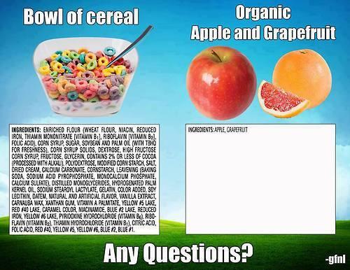
Ok look. This is nuts. Are fruit loops healthier than an actual piece of fruit? Pretty much no, never. Individual quirks of metabolism aside, I can’t think of a single reason I would ever tell someone to eat fruit loops over actual fruit. Every time I see this it’s like a smart off to say stupid shit. I guarantee I do more biochem on a regular basis than most (and you can ask other docs that know me, like @notacleverturnip, I do so much biochem all the time). I don’t give a fuck about pronunciation, I’m just trying to stop people from getting diabetes. The chemical scare tactic is not particularly useful but neither is telling someone that the ingredient list on fruit loops is like the same fucking thing as an apple. I swear to the gods of biochem and nutrition if this post (in all of its iterations, I don’t care for the scare tactics either) comes around again I’m going to throw a blog tantrum. Again.If you look at the ingredients list and it’s a bunch of words you don’t even know… neither does your body (x)
Just like if you break apples and grapefruit down into their chemical components, I’m willing to bet that most people wouldn’t recognize the “ingredients” either. It’s a bunch of words you don’t even know:
External imageDon’t use these scare tactics - Chemicals aren’t inherently bad. Literally everything is made up chemicals. Trust me, your body knows what niacin is. It knows how to digest fructose and calcium sulfate. Even if you only consume the most basic and “real” foods that are pulled directly off the vine, you’re still ingesting a series of chemical compounds that you probably can’t pronounce. That’s okay.
thanks to drhoz for submitting!
“If you can’t pronounce it, it’s bad for you” is literally the worst pseudo-scientific scaremongering bullshit tactic. I hate it so much.
I’m pretty sure you can pronounce “arsenic”, but that doesn’t change the fact that arsenic is highly toxic. On the other hand, you couldn’t pronounce “cycloadenosine monophosphate” or “nicotine-amide-dinucleotide-phosphate”, though both of them serve vital roles in human biochemistry and you would die if your body wouldn’t produce them.
Cyanide: Easy to pronounce, very bad for you.
Eicosapentaenoic acid: Difficult to pronounce, very good for you.
It’s more important to know what the chemicals are and why they’re in there. Anti-intellectualism helps no one.
– James Kennedy, ‘Chemophobia’ is irrational, harmful – and hard to break
I’m gonna keep reblogging this until my knuckles fall off.
This is especially hilarious because grapefruit is well known for being dangerous for some people because of how it can interact with certain medications. Do fruit loops do that?
“Poison is in everything, and no thing is without poison. The dosage makes it either a poison or a remedy.” - Paracelsus
Fava beans are like *known* to fuck up certain people with G6PD deficiency - which is insanely common.
No one is saying fruit loops are healthy or the same as an apple. They’re saying you shouldn’t make eating decisions based on whether you can pronounce the ingredients or not. There’s obviously way more to nutrition than that, but fruit loops aren’t dangerous just because the ingredients are difficult to pronounce. That’s literally all this post is trying to say. It’s by no means a recommendation that fruit loops are super great and healthy.
-
Vision Guessed
What do you see? The answer lies in the eye of the beholder. In this case, quite literally. The image, taken by Kim Baxter at Cambridge University Hospitals NHS Foundation Trust and a 2017 Wellcome Image Awards, depicts blood vessels feeding the retina of a human eye. They appear as white, spidery lines due to a fluorescent dye passing through.
-
New Assay May Lead to Better Treatment for Rheumatoid Arthritis
Researchers at NYU Langone Medical Center have developed a test to measure the immunologic defect that triggers the inflammation present in rheumatoid arthritis. They believe that clinical trials for new rheumatoid arthritis drugs should shift from their sole focus on relieving inflammation to eliminating the B cells that produce the antibodies that cause this defect.
“We have developed a test for measuring the underlying autoimmunity in rheumatoid arthritis patients that should be used to evaluate new treatment regimens,” says senior author Gregg Silverman, MD, professor in the departments of Medicine and Pathology at NYU Langone and co-director of its Musculoskeletal Center of Excellence. “We believe this provides a road to a cure for rheumatoid arthritis.”
Funding: This work was supported by the National Institutes of Health (NIH), an American Recovery and Reinvestment Act supplement, and a National Institute of Allergy and Infectious Diseases/NIH and NYU School of Medicine-Immunology and Inflammation Training grant.
Raise your voice in support of expanding federal funding for life-saving medical research by joining the AAMC’s advocacy community.
-
An Unexpected New Lung Function Has Been Found - They Make Blood:
Researchers have discovered that the lungs play a far more complex role in mammalian bodies than we thought, with new evidence revealing that they don’t just facilitate respiration - they also play a key role in blood production.
In experiments involving mice, the team found that they produce more than 10 million platelets (tiny blood cells) per hour, equating to the majority of platelets in the animals’ circulation. This goes against the decades-long assumption that bone marrow produces all of our blood components.
Researchers from the University of California, San Francisco also discovered a previously unknown pool of blood stem cells that makes this happen inside the lung tissue - cells that were incorrectly assumed to mainly reside in bone marrow.
“This finding definitely suggests a more sophisticated view of the lungs - that they’re not just for respiration, but also a key partner in formation of crucial aspects of the blood,” says one of the researchers, Mark R. Looney.
“What we’ve observed here in mice strongly suggests the lung may play a key role in blood formation in humans as well.”
While the lungs have been known to produce a limited amount of platelets - platelet-forming cells called megakaryocytes have been identified in the lungs before - scientists have long assumed that most of the cells responsible for blood production are kept inside the bone marrow.
Here, a process called haematopoiesis was thought to churn out oxygen-laden red blood cells, infection-fighting white blood cells, and platelets - blood components required for the clotting that halts bleeding.
But scientists have now watched megakaryocytes functioning from within the lung tissue to produce not a few, but most of the body’s platelets.
-
Neuroscientists Have Accidentally Discovered a Whole New Role for the Cerebellum:
One of the best-known regions of the brain, the cerebellum accounts for just 10 percent of the organ’s total volume, but contains more than 50 percent of its neurons.
Despite all that processing power, it’s been assumed that the cerebellum functions largely outside the realm of conscious awareness, instead coordinating physical activities like standing and breathing. But now neuroscientists have discovered that it plays an important role in the reward response - one of the main drives that motivate and shape human behaviour.
Not only does this open up new research possibilities for the little region that has for centuries been primarily linked motor skills and sensory input, but it suggests that the neurons that make up much of the cerebellum - called granule cells - are functioning in ways we never anticipated.
“Given what a large fraction of neurons reside in the cerebellum, there’s been relatively little progress made in integrating the cerebellum into the bigger picture of how the brain is solving tasks, and a large part of that disconnect has been this assumption that the cerebellum can only be involved in motor tasks,” says one of the team, Mark Wagner, from Stanford University.
“I hope that this allows us to unify it with studies of more popular brain regions like the cerebral cortex, and we can put them together.”
-
For the first time, Tübingen neuroscientists were able to differentiate between active and inactive cells in the brain morphologically, i.e. based on the cells’ structure. Investigating granule cells in the rat’s brain, they found a much larger proportion of inactive than active cells.
Many things we think we know about the world have their origin in popular culture, not science. The most well-known false ‘fact’ about the brain is the misconception that we only use ten percent of the brain’s overall capacity. This so-called ’ten percent myth’, while accepted as such by neuroscientists, still regularly figures in advertisement, but also in books and short stories as well as films. As with any myth, however, there is a kernel of truth at the core of the matter: many neurons remain dormant for most if not all of our life, even while their direct neighbours show regular activity.
A team of neuroscientists led by Dr. Andrea Burgalossi of the Werner Reichardt Centre for Integrative Neuroscience (CIN) at the University of Tübingen have now taken an important step towards understanding why some neurons are active and others are not: they can tell them apart morphologically. To be able to do so, the investigators employed so-called juxtacellular recordings in freely-moving rats. With this technique, electrodes are inserted right next to individual, functioning neurons in live organisms. This allows recording action potentials from these neurons while they work, and while simultaneously identifying the cells that the recordings are taken from for later analysis.
During this analysis, morphological traits of the analysed cells are identified, most importantly their dendritic arbors, i.e. the filament structures which receive input signals from other neurons. The cells under investigation were granule cells (GCs) in the rat’s dentate gyrus (DG). Dentate GCs have been shown to be intimately connected to individual memories of places and individuals, and thus playing a central role in memory tasks.
The researchers recorded from 190 GCs, only 27 of which they found to be active (ca. 14 percent). While this seems to give credibility to the ‘ten percent myth’, the team actually expected this outcome, as the DG is a brain structure where in any given task, only a very small percentage of neurons take part, while their neighbours remain dormant, waiting for their ‘cue’, as it were. Memory functions in the brain work according to a principle that neuroscientists call ‘sparse coding’, i.e. a comparatively small number of neurons encode complex information – possibly to make overlap between different memories more unlikely.
Using a smaller subsample, the scientists looked for correlations between active and passive functionality and the respective cells’ morphology. Their results show that active GCs have much more complex dendritic arbors. They not only transfer and receive information from many more neurons than the inactive ones, they also have better cellular ‘infrastructure’ to do so. Despite their as of yet limited sampling, the scientists are positive that they can now tell apart active and inactive GCs, mostly by merely looking at them. “Explaining the causes of activity in some and inactivity in other neurons may still take a long time”, cautions Burgalossi, leader of the research group. “But finding a direct link between function and morphology is an important step forward. It will be even more challenging to find evidence of causality. But we are on the right track.”
-
Researchers Develop “MAGIC Algorithm” to Predict Whether Bone Marrow Transplant Patients May Die From Common Complication
Researchers at Mount Sinai Health System have discovered a way to predict whether blood cancer patients who received a bone marrow transplant will develop graft-versus-host disease, a common and often lethal complication.
The study, which involved 11 cancer centers internationally, used blood samples from almost 1,300 bone marrow transplant patients and found that two proteins (ST2 and REG3a) present in blood drawn a week after a transplant can predict whether a patient will develop a lethal version of graft-versus-host disease. Scientists at the Mount Sinai Acute GVHD International Consortium (MAGIC) created an algorithm, dubbed the “MAGIC algorithm,” that determines a patient’s risk of developing the disease by measuring concentrations of these proteins.
The research was published in JCI (The Journal of Clinical Investigation) Insight.
“The MAGIC algorithm gives doctors a roadmap to save many lives in the future. This simple blood test can determine which bone marrow transplant patients are at high risk for a lethal complication before it occurs,” says James L.M. Ferrara, MD, Professor of Pediatrics, Oncological Sciences and Medicine, Hematology and Medical Oncology at The Tisch Cancer Institute at the Icahn School of Medicine at Mount Sinai, and Co-director of MAGIC. “It will allow early intervention and potentially save many lives.”
Funding: The study was supported by grants P01 CA03942 and P30 CA106521 from the National Cancer Institute, an American Cancer Society Clinical Research Professorship (to Dr. Ferrara) and a Doris Duke Charitable Foundation Clinical Research Mentorship.
Raise your voice in support of expanding federal funding for life-saving medical research by joining the AAMC’s advocacy community.
-
Neuroscientists call for deep collaboration to ‘crack’ the human brain
The time is ripe, the communication technology is available, for teams from different labs and different countries to join efforts and apply new forms of grassroots collaborative research in brain science. This is the right way to gradually upscale the study of the brain so as to usher it into the era of Big Science, claim neuroscientists in Portugal, Switzerland and the United Kingdom. And they are already putting ideas into action.
In a Comment in the journal Nature, an international trio of neuroscientists outlines a concrete proposal for jump-starting a new, bottom-up, collaborative “big science” approach to neuroscience research, which they consider crucial to tackle the still unsolved great mysteries of the brain.
How does the brain function, from molecules to cells to circuits to brain systems to behavior? How are all these levels of complexity integrated to ultimately allow consciousness to emerge in the human brain?
The plan now proposed by Zach Mainen, director of research at the Champalimaud Centre for the Unknown, in Lisbon, Portugal; Michael Häusser, professor of Neuroscience at University College London, United Kingdom; and Alexandre Pouget, professor of neuroscience at the University of Geneva, Switzerland, is inspired by the way particle physics teams nowadays mount their huge accelerator experiments to discover new subatomic particles and ultimately to understand the evolution of the Universe.
“Some very large physics collaborations have precise goals and are self-organized”, says Zach Mainen. More specifically, his model is the ATLAS experiment at the European Laboratory of Particle Physics (CERN, near Geneva), which includes nearly 3,000 scientists from tens of countries and was able (together with its “sister” experiment, CMS) to announce the discovery of the long-sought Higgs boson in July 2012.
Although the size of the teams involved in neuroscience may not be nearly comparable to the CERN teams, the collaborative principles should be very similar, according to Zach Mainen. “What we propose is very much in the physics style, a kind of ‘Grand Unified Theory’ of brain research, he says. “Can we do it? Clearly, it’s not going to happen within five years, but we do have theories that need to be tested, and the underlying principles of how to do it will be much the same as in physics.”
To help push neuroscience research to take the leap into the future, the three neuroscientists propose some simple principles, at least in theory: “focus on a single brain function”; “combine experimentalists and theorists”; “standardize tools and methods”; “share data”; “assign credit in new ways”. And one of the fundamental premises to make this possible is to “engender a sphere of trust within which it is safe [to share] data, resources and plans”, they write.
Needless to say, the harsh competitiveness of the field is not a fertile ground for this type of “deep” collaborative effort. But the authors themselves are already putting into practice the principles they advocate in their article.
“We have a group of 20 researchers (10 theorists and 10 experimentalists), about half in the US and half in the UK, Switzerland and Portugal” says Zach Mainen. The group will focus on only one well-defined goal: the foraging behavior for food and water resources in the mouse, recording activity from as much of the brain as possible - at least several dozen brain areas.
“By collaboration, we don’t mean business as usual; we really mean it”, concludes Zach Mainen. “We’ll have 10 labs doing the same experiments, with the same gear, the same computer programs. The data we will obtain will go into the cloud and be shared by the 20 labs. It’ll be almost as a global lab, except it will be distributed geographically.”
-
A SISSA research study published in a special issue of the journal Brain and Cognition, completely dedicated to the cognitive neuroscience of food, analyzes the lexical-semantic deficits of the food category in patients suffering from neurodegenerative diseases like Alzheimer’s. The study shows that knowledge about food is preserved more than other categories of stimuli, even in the case of severe syndromes. Further, perception of caloric intake affects a person’s ability to remember the name of a food; the higher the calories, the more knowledge is preserved. Professor Raffaella Rumiati of the International School for Advanced Studies (SISSA) in Trieste, first author and expert in semantic categorization of food, also served as editor of the special issue (along with Giuseppe Di Pellegrino, University of Bologna), and wrote the introduction to the issue.
Perhaps it is because it is so crucial to our survival that lexical and semantic knowledge related to food is relatively well preserved even in diseases that lead to a general decline in memory and cognition, such as Alzheimer’s and Aphasia Primary Progressive. Raffaella Rumiati and her team at SISSA, in collaboration with Caterina Silveri of Catholic University “Agostino Gemelli” in Rome, observed the phenomenon while testing the cognitive performance of two groups of patients and a control group of healthy people in tasks concerning visual recognition of food and comprehension.
“It should not be surprising that food resists even generalized cognitive decline,” says Rumiati. “It is not difficult to imagine how evolutionary pressure could lead to increased strength in cognitive processes related to fast recognition of what is probably the most important stimulus for survival.” Another fact revealed by the study supporting food supremacy was that in all three groups, patients and control, food information was processed better than “non–food.” Adds Rumiati, “We know from the literature that the names of the most caloric foods are acquired early in life.”
Rumiati and colleagues discovered another interesting detail: the perception of caloric intake of each food is proportional to the strength with which we recognise their names. The more caloric the food seems, the better it is preserved. “This phenomenon may be closely related to what I said earlier: the more nutritious the food, the more important it is to recognize it.”
-
Researcher Reveals Clues to Immunity as a Cause of High Blood Pressure
A University of Arkansas for Medical Sciences (UAMS) researcher has shed light on the role of immune cells inside the kidneys in the development of salt-sensitive high blood pressure, publishing his findings in Nature Communications.
“High blood pressure is very common, and this salt-sensitive version is present in about 40-50 percent of cases of hypertension initially and becomes worse as the disease progresses,” said Shengyu Mu, Ph.D., assistant professor in the Department of Pharmacology and Toxicology in the UAMS College of Medicine. “For many years, the recommendation for heart health has been to eat less salt. But studies have shown that while it does lower blood pressure; too little salt also increases the rate of cardiovascular events and mortality in patients, which is all the more reason for us to develop a better understanding of the causes of salt-sensitivity.”
Specifically, Mu’s work uncovered the interaction of a particular type of white blood cell with kidney cells. Scientists suspected that these cells – T lymphocytes, or T cells –demonstrating that too many T cells in the kidneys might be the cause of salt sensitivity of high blood pressure.
Funding: This study was supported by American Heart Association Beginning Grant-in-Aid and financial support from Dr. Philip Palade and the University of Arkansas for Medical Sciences Foundation. The researchers are also funded in part by the National Institutes of Health and a VA Merit Award.
Raise your voice in support of expanding federal funding for life-saving medical research by joining the AAMC’s advocacy community.
-
Epstein-Barr virus and cancer: New tricks from an old dog
After an infection with the Epstein-Barr virus (EBV), the virus persists in the body throughout a person’s lifetime, usually without causing any symptoms. About one third of infected teenagers and young adults nevertheless develop infectious mononucleosis, also known as glandular fever or kissing disease, which usually wears off after a few weeks. In rare cases, however, the virus causes cancer, particularly lymphomas and cancers of the stomach and of the nasopharynx.
Scientists have been trying for a long time to elucidate how the viruses reprogram cells into becoming cancer cells. “The contribution of the viral infection to cancer development in patients with a weakened immune system is well understood” says Henri-Jacques Delecluse, a cancer researcher at the German Cancer Research Center (Deutsches Krebsforschungszentrum, DKFZ) in Heidelberg. “But in the majority of cases, it remains unclear how an EBV infection leads to cancer development.”
In their present publication, Delecluse, in collaboration with Ingrid Hoffmann, also from the DKFZ, and their respective groups present a new and surprising explanation for this phenomenon. The scientists have shown for the first time that a protein component of the virus itself promotes the development of cancer. When a dividing cell comes in contact with Epstein-Barr viruses, a viral protein present in the infectious particle called BNRF1 frequently leads to the formation of an excessive number of spindle poles (centrosomes). As a result, the chromosomes are no longer divided equally and accurately between the two daughter cells – a known and acknowledged cancer risk factor. By contrast, Epstein-Barr viruses that had been made deficient of BNRF1 did not interfere with chromosome distribution to the daughter cells.
Anatoliy Shumilov, Ming-Han Tsai, Yvonne T. Schlosser, Anne-Sophie Kratz, Katharina Bernhardt, Susanne Fink, Tuba Mizani, Xiaochen Lin, Anna Jauch, Josef Mautner, Annette Kopp-Schneider, Regina Feederle, Ingrid Hoffmann, Henri-Jacques Delecluse. Epstein–Barr virus particles induce centrosome amplification and chromosomal instability. Nature Communications, 2017; 8: 14257 DOI: 10.1038/ncomms14257
After an infection with the Epstein-Barr virus (EBV), the virus persists in the body throughout a person’s lifetime. Credit: © Henri-Jacques Delecluse/DKFZ
-
Bed Bugs: The Worst Bed Pest and How To Get Rid of Them
Bed bugs have been and are still one of the worst nightmares of every homeowner. Can you imagine that creepy crawlies sleeping with you and squeaking every night? But fortunately, there are already proven effective methods to prevent and handle home pestilence from these viruses without spraying the entire house with harmful and toxic chemicals.
Despite the usual effective sprays, repellents, and traps, these can sometimes be hazardous to you if not lethal when choked or even touched. Laying open to these preventive methods can cause eye, skin and respiratory irritation and even cause cancer when mishandled. But before we go ahead and discuss the steps on how to get rid of bed bugs, what are bed bugs, anyway?
KNOWING YOUR REAL ENEMY
BED BUGS
What They Look Like – a brown or a reddish brown animal that has a flat, oval body and the same as the size of an apple seed.
Headquarters – usually, they hang out around and in the bed itself. Their small and flat body makes it easy for them to hide around the corner of the bed frames, headboards, mattresses, behind your room wallpaper and even your clothes.
Danger – this scourge does not transmit or pass through diseases. They only suck human and animal blood. Their bites cause itchiness and swelling. The person dealing with the pestilence can be both anxious and may experience insomnia.
Good thing, we have some alternative ways on how to prevent and stop bed bugs. Methods that are milder, healthier and more environment-friendly.
HOW CAN I KEEP BED BUGS OUT?
The real horror of bugs in the bedroom can never be understood unless you experience it. Here are a few effective steps on how to KEEP THEM OUT:
Clean up – Every virus loves a dirty environment. Clean up spills and crumbs right away every after bedroom meal.
Stay dry – Wipe any cold water sweat from a glass on your bedside table.
Heat up the textiles – all the linens (bed sheets, comforter, pillow cases, blankets, curtains and even towels) where bugs might hide should be washed with the highest temperature available for at least thirty minutes.
Scrub the mattress – this ensures that the bed bugs in the mattress are also taken care off. It’s not enough to just wash the linens without touching the mattress. Make sure you use something like a stiff brush and a vacuum.
Decide if the mattress can still be kept – there are times when the mattress will no longer be useful due to the huge amount of bed bugs on it. If that’s the case, unfortunately, you already need to replace your bed mattress. Be careful in throwing buggy materials. Properly wrap it and label it as a material with bugs.
TAKEAWAY
Most of the time, we think of our room as our own comfort zone. And it is very important that you’re comfortable in it. That’s why it is very important to clean our room every now and then. Never ignore that itchy feeling you get whenever you lay down in your bed. It might be those annoying blood-sucking bed bugs waiting to attack you.
-
One of the worst epidemics in human history, a sixteenth-century pestilence that devastated Mexico’s native population, may have been caused by a deadly form of salmonella from Europe, a pair of studies suggest.
In one study, researchers say they have recovered DNA of the stomach bacterium from burials in Mexico linked to a 1540s epidemic that killed up to 80% of the country’s native inhabitants. The team reports its findings in a preprint posted on the bioRxiv server on 8 February.
This is potentially the first genetic evidence of the pathogen that caused the massive decline in native populations after European colonization, says Hannes Schroeder, an ancient-DNA researcher at the Natural History Museum of Denmark in Copenhagen who was not involved in the work. “It’s a super-cool study.”
In 1519, when forces led by Spanish conquistador Hernando Cortés arrived in Mexico, the native population was estimated at about 25 million. A century later, after a Spanish victory and a series of epidemics, numbers had plunged to around 1 million. Read more.
-
Fibrodysplasia Ossificans Progressiva (FOP) is a congenital condition that literally turns the connective tissue of those who suffer from it into bone, slowly seizing up their joints until finally they’re trapped inside their own body.
The bone growth is aggravated by damage to the soft tissue confounded by the fact that early symptoms look like fibrosis or cancer, coupled with the rarity of FOP means that doctors usually prescribe a biopsy which only hastens the bone growth.
Currently there is no known cure as removal of the growths only results in the body repairing the surrounding tissue with more bone, strangely the diaphragm and heart tissue is spared leaving sufferers condemned to a slow, tortuous existence.
-
Image SR6169 (Balancing stone from inner ear, SEM) ©Susumu Nishinaga/Science Source
Image DA8958 (Acoustic Macula) ©BSIP/Science Source
Image DA8959 (Acoustic Macula) ©BSIP/Science Source
I’ve Got Rocks In My Head…
…and so do all of you. The image at the top is a colored scanning electron micrograph (SEM) of crystals of calcium carbonate on the surface of an otolith. They are the “balancing stones” of the inner ear and are found in our Acoustic Macula.
See more images of the Acoustic Macula
The acoustic macula is responsible for our static equilibrium (position of the head) and participates in dynamic equilibrium (recognition of the linear accelerations). Located at the level of the inner ear, the macula is composed of hair cells (in orange), constituting the sensorial receptors, and of supporting cells (in pink). Each hair cell possesses between 40 to 70 stereocilia and a single kinocilium.
See SEMs of Inner Ear Hair Cells
The supporting cells secrete a gelatinous substance forming the otolithic membrane, in which embed the stereocilia and kinocilia. This membrane is covered with a layer of those calcium carbonate crystals (shown at top). Each hair cell forms a synapse with a sensitive neuron (in yellow) and a motor neuron (in green) of the vestibular branch of the auditory nerve.
See more images of the Inner Ear
During a sharp acceleration leading the head forward (during the ascension in the roller coaster, for example), the inertia causes a sliding movement backwards of the otolithic membrane and the otoliths, that move the stereocilia and kinocilia with them. This leads to a stimulation of the vestibular nerve, enabling the recognition of the movement.
What would a trip to an amusement park be without your inner ear?!
All images © Science Source
-
Where is My Mind? New Study Looks for the Cortical Conscious Network
Our brain is a very complex network, with approximately 100 billion neurons and 100 trillion synapses between the neurons. In order to cope with its enormous complexity, and understand how the brain works and eventually forms our conscious mind, science uses advanced mathematical tools. Ultimately, scientists seek to understand how a global phenomenon such as consciousness can emerge from our neuronal network.
A team of Bar-Ilan University physicists, led by Prof. Shlomo Havlin and Prof. Reuven Cohen, used network theory in order to cope with this complexity and to determine how the structure of the human cortical network can support complex data integration and conscious activity. The gray area of the human cortex, the neuron cell bodies, were scanned with MRI imaging and used to form 1,000 nodes in the cortical network. The white matter of the human cortex, the neuron bundles, were scanned with DTI imaging, forming 15,000 links or edges which connected the network’s nodes. In the end of this process, their network was an approximation of the structure of the human cortex.
Their research, recently published in New Journal of Physics, attempts to decompose the structural layers of the cortical network to different hierarchies, enabling the identification of the network’s nucleus from which our consciousness could emerge.
Previous studies have shown that the human cortex is a network with small world properties, which means that it has many local structures and some shortcuts from global structures which connect faraway areas (similar to the difference between local buses and cross-country trains). The cortex also has many hubs, which are nodes that have a high number of links (like central stations), that are also strongly interconnected between themselves, making it easy to travel between the brain’s information highways.
According to Nir Lahav, the lead author of the study, “In order to examine how the structure of the network can support global emerging phenomena, like consciousness, we applied a network analysis called k-shell decomposition. This analysis takes into account the connectivity profile of each node making it easy to uncover different neighborhoods of connections in the cortical network, we called shells.” The most connected neighborhood in the network is termed the network’s nucleus. Lahav explains, “In the process we peel off different shells of the network to get the most connected area of the network, the nucleus. Until today scientists were only interested in the network’s nucleus, but we found that these different shells can hold important information about how the brain integrates information from the local levels of each node to the entire global network. For the first time we can build a comprehensive topological model of our cortex.”
This topological model reveals that the network’s nucleus includes 20% of all nodes and that the remaining 80% are strongly connected across all the different shells. Interestingly, when comparing this topology to that of other networks, such as the internet, some noticeable differences can be seen. For instance, in internet network topology almost 25% of the nodes are isolated, meaning they don’t connect to any other shells but the nucleus. In the cortical network, however, there are hardly any isolated nodes. It seems that the cortex is much more connected and efficient than the internet.
Looking at all the different shells of the cortical network, the authors were able to define the network’s hierarchical structure and essentially model how information flows within the network. The structure revealed how shells of low connectivity are nodes that typically perform specific functions like face recognition. From there the data are transferred to higher, more connected shells that enable additional data integration, and there regions of the executive network and working memory can be seen. With these areas we can focus on task performance, for example.
The integrated information then ‘travels’ to the most connected neighborhood of nodes, the nucleus, which spans across several regions of the cortex. According to Lahav, “It’s an interconnected collective which is densely linked with itself and can perform global functions due to its great amount of global structures that are widespread across the brain.”
Which global function might the nucleus serve? The authors suggest the answer is no less than consciousness itself.
“The connection between brain activity and consciousness is still a great mystery,” says Lahav. The main hypothesis today is that in order to create conscious activity, the brain must integrate relevant information from different areas of the network. According to this theory, led by Prof. Giulio Tononi, from the University of Wisconsin, if the level of integrated information crosses a certain limit, a new and emergent state is entered, consciousness. This model suggests that consciousness depends on both information integration and information segregation. Loosely speaking, consciousness is generated by a “central” network structure with high capacity for information integration, with the contribution of sub-networks that contain specific and segregated information, without being part of the central structure. In other words, certain parts of the brain are more involved than others in what we can call the conscious complex of the brain, yet other connected parts still contribute, working quietly outside the conscious complex.
The authors demonstrate how the nucleus and the different shells satisfy all of the requirements of these recent consciousness theories. The different shells calculate and contribute to data integration without actually being part of the conscious complex, while the nucleus receives relevant information from all other hierarchies and integrates it to a unified function using its global interconnected structure. The nucleus could thus be this conscious complex, serving as a platform for consciousness to emerge from the network activity.
When the authors examined the different regions that make up the nucleus they revealed that, indeed, these regions have been previously associated with conscious activities. For example, structures within the brain’s midline, which form the majority of the network’s nucleus, were found to be associated with the stream of consciousness and some research, like that of Prof. Georg Northoff from the University of Ottawa, have suggested that these regions are involved with creating our sense of self.
“Now we need to use this analysis on the whole brain and not only on the cortex in order to reveal a more exact model of the brain’s hierarchy, and later on to try to understand what exactly are the neuronal dynamics that lead to such global integration and, ultimately, consciousness,“ says Lahav.
“Profound questions need a profound answer that can usually be found only in physics. Physics tries to uncover the basic laws of nature by constructing general mathematical equations that can describe as many natural phenomena as possible. These mathematical equations reveal fundamental aspects of reality. If we really want to understand what consciousness is and how the brain works, we have to develop the mathematical equations of our brain and our conscious mind. We are not there yet, in fact we are quite far away from this goal, but I feel that this should be our ‘holy grail’ and we have already begun the process of getting there,” he adds.


currentsinbiology: Folk contraceptives lead researchers to drugs...
in Topics
Posted
currentsinbiology:
Via